
File:Polychaeta anatomy en.svg

Size of this PNG preview of this SVG file: 800 × 475 pixels. Other resolutions: 320 × 190 pixels | 640 × 380 pixels | 1,024 × 608 pixels | 1,280 × 760 pixels | 2,560 × 1,520 pixels | 1,999 × 1,187 pixels.
Original file (SVG file, nominally 1,999 × 1,187 pixels, file size: 297 KB)
| Photographer | |
| Description |
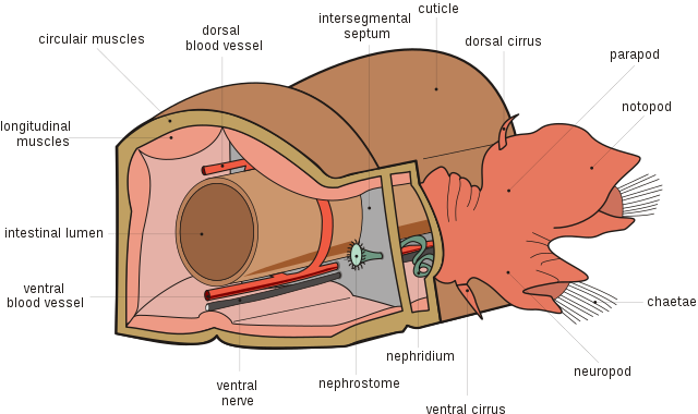
 English: Section of two segments of a Polychaete (Nephtys sp.), showing internal anatomy. The large structure on the right is a parapod. (labeled in English)
Français : Schéma d'une coupe transversale d'un polychète (Nephtys sp.). On peut observer un parapode sur la droite. (Texte en Anglais)
Nederlands: Engelstalige interne anatomie van een borstelworm (Nephtys sp.) a.d.h. van een doorsnede door twee segmenten. Aan de rechterzijde ziet u een parapode.
Português: Secção de dois segmentos de Polychaeta (Nephtys sp.), mostrando a anatomia interna. |
| Date | 12 February 2007 |
| Source | Own work |
| Permission (Reusing this file) |
This file is licensed under the Creative Commons Attribution-Share Alike 4.0 International license. Attribution: © Hans Hillewaert
|
| Other versions |
[]
|
| Other licenses | More (4100+) and new images on : High resolution files on request. . |
| Attribution (required by the license) | © Hans Hillewaert / |
|
This image was selected as picture of the day on Wikimedia Commons for 18 November 2009. It was captioned as follows: English: Section of two segments of a Polychaete (Nephtys sp.), showing internal anatomy. The large structure on the right is a parapod. (Labeled in English.) Other languages:
Bosanski: Presjek dva segmenta Polychaete (rod Nephtys), koji pokazuje unutrašnju anatomiju. Velika struktura desno je parapodij. Čeština: Řez dvěma články mnohoštětinatce (Nephtys sp.); velký útvar vpravo je parapodium (popisky v angličtině, dostupná je i česká verze) Dansk: Snit gennem to led af en havbørsteorm (Nephtys), som viser dens indre anatomi. (Engelske tekster) English: Section of two segments of a Polychaete (Nephtys sp.), showing internal anatomy. The large structure on the right is a parapod. (Labeled in English.) Español: Sección de dos segmentos de poliqueto (Nephtys sp.) mostrando la anatomía interna. La estructura grande de la derecha es un parápodo. (Rótulos en inglés.) Français : Schéma d'une coupe transversale d'un polychète (Nephtys sp.). On peut observer un parapode sur la droite. (Texte en Anglais) Magyar: Soksertéjűek vázlatos felépítése, a jobboldali lebeny az evező (parapódium) Nederlands: Engelstalige doorsnede door twee segmenten van de interne anatomie van een Nephtys sp. (een borstelwormsoort uit de familie Nephtyidae van de orde Aciculata uit de onderklasse Palpata). Aan de rechterzijde is een parapode (uitsteeksel dat dient als kieuw en de chitinen chaeta (borstels) bevat) zichtbaar. Polski: Schemat budowy wieloszczetów na podstawie dwóch segmentów Nephtys sp. Po prawej dobrze widoczne parapodium. Português: Secção de dois segmentos de Polychaeta (Nephtys sp.), mostrando a anatomia interna. Македонски: Пресек на два дела од Полихет (Nephtys sp.) со приказ на внатрешната анатомија. Големиот дел од лево е параподија. বাংলা : অন্তঃস্থ অঙ্গসংস্থান নির্দেশিত দ্বিপর্ব বিশিষ্ট একটি পলিকেটের ব্যবচ্ছেদ চিত্র। ডান পাশের বড়ো অংশটি হচ্ছে প্যারাপড (চিত্রটি ইংরেজিতে চিহ্নিত হয়েছে)। 中文: 多毛纲生物的2段体节的解剖图解,右侧的部分为疣足 中文(繁體): 多毛綱生物的2段體節的解剖圖解,右側的部分為疣足 |
This W3C-unspecified vector image was created with Inkscape .

|
This SVG file contains embedded text that can be translated into your language, using any capable SVG editor, text editor or the SVG Translate tool. For more information see: About translating SVG files. |
Labeling
| English | Nederlands | Espagnol | Português |
|---|---|---|---|
| circular muscles | circulaire spieren | Músculo circular | Músculo circular |
| dorsal blood vessel | dorsaal bloedvat | Vaso dorsal | Vaso dorsal |
| intersegmental septum | intersegmentair septum | Septo intersegmentario | Septo Metamérico |
| cuticle | cuticula | Cutícula | Cutícula |
| dorsal cirrus | dorsale cirrus | Cirro dorsal | Tubo dorsal |
| parapod | parapode | Parápodo | Parápode |
| notopod | notopodium | Notopodio | Notopode |
| chaetae | chaetae | Quetas | cerdas |
| neuropod | neuropodium | Neuropodio | Neuropode |
| ventral cirrus | ventrale cirrus | Cirro ventral | Tubo ventral |
| nephridium | nephridium | Nefridio | Nefrídio |
| nephrostome | nephrostoom | Nefrostoma | Nefróstoma |
| ventral nerve | ventrale zenuw | Nervio ventral | Nervo ventral |
| ventral blood vessel | ventraal bloedvat | Vaso ventral | Vaso ventral |
| intestinal lumen | darmholte | Intestino | Intestino |
| longitudinal muscles | longitudinale spieren | Músculo longitudinal | Músculo longitudinal |
File history
Click on a date/time to view the file as it appeared at that time.
| Date/Time | Thumbnail | Dimensions | User | Comment | |
|---|---|---|---|---|---|
| current | 20:29, 18 April 2020 | | 1,999 × 1,187 (297 KB) | Nestor pamir | File uploaded using svgtranslate tool (https://tools.wmflabs.org/svgtranslate/). Added translation for ca. |
| 20:27, 18 April 2020 |  | 1,999 × 1,187 (295 KB) | Nestor pamir | File uploaded using svgtranslate tool (https://tools.wmflabs.org/svgtranslate/). Added translation for ca. | |
| 07:02, 12 February 2007 |  | 1,999 × 1,187 (292 KB) | Lycaon | {{Information |Description= Diagram of Polychaeta anatomy in English |Source= Self |Date= 12 02 2007 |Author= Hans Hillewaert Lycaon |Permission= {{self|cc-by-sa-2.5}} |other_versions=Image:Polychaeta anatomy nl.svg }} {{Translation |
File usage
The following 5 pages use this file:
Global file usage
The following other wikis use this file:
- Usage on bg.wikipedia.org
- Usage on bn.wikipedia.org
- Usage on da.wikipedia.org
- Usage on en.wikipedia.org
- Usage on hr.wikipedia.org
- Usage on is.wikipedia.org
- Usage on ja.wikipedia.org
- Usage on ko.wikipedia.org
- Usage on os.wikipedia.org
- Usage on outreach.wikimedia.org
- Usage on pam.wikipedia.org
- Usage on pl.wikipedia.org
- Usage on pt.wikipedia.org
- Usage on ru.wikipedia.org
- Usage on ru.wikinews.org
- Usage on sh.wikipedia.org
- Usage on www.wikidata.org
- Usage on zh.wikipedia.org