
File:Anatomy of the Human Ear-Number.svg

Size of this PNG preview of this SVG file: 674 × 592 pixels. Other resolutions: 273 × 240 pixels | 547 × 480 pixels | 874 × 768 pixels | 1,166 × 1,024 pixels | 2,332 × 2,048 pixels.
Original file (SVG file, nominally 674 × 592 pixels, file size: 98 KB)
Summary
| Description |
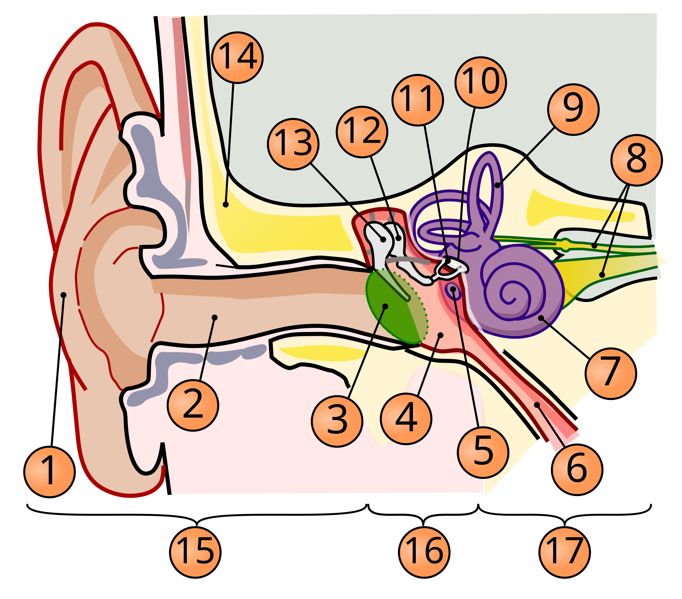
English: A diagram of the anatomy of the human ear.
Legend: Brown is outer ear.
Red is middle ear.
Purple is inner ear. |
||
| Date | (UTC) | ||
| Source | |||
| Author |
|
||
| Other versions |
[]
|
||
| SVG development | This diagram was created with Inkscape. This diagram uses embedded text that can be easily translated using a text editor.
|


Licensing
I, the copyright holder of this work, hereby publish it under the following license:
This file is licensed under the Creative Commons Attribution 2.5 Generic license.
- You are free:
- to share – to copy, distribute and transmit the work
- to remix – to adapt the work
- Under the following conditions:
- attribution – You must give appropriate credit, provide a link to the license, and indicate if changes were made. You may do so in any reasonable manner, but not in any way that suggests the licensor endorses you or your use.
Original upload log
This image is a derivative work of the following images:
- File:Anatomy_of_the_Human_Ear.svg licensed with Cc-by-2.5
- 2009-04-28T21:49:54Z Mike.lifeguard 800x600 (91972 Bytes) Malleus and incus were swapped O.o
- 2009-02-15T14:59:07Z Inductiveload 800x600 (91617 Bytes) added tympanic cavity
- 2009-02-15T14:50:03Z Inductiveload 800x600 (89435 Bytes) {{Information |Description={{en|1=A diagram of the anatomy of the human ear.}} |Source=[[:File:10.1371_journal.pbio.0030137.g001-L.jpg]], vectorised by [[User:Inductiveload|Inductiveload]] |Author=[[User:Inductiveload|Induct
Uploaded with derivativeFX
File history
Click on a date/time to view the file as it appeared at that time.
| Date/Time | Thumbnail | Dimensions | User | Comment | |
|---|---|---|---|---|---|
| current | 16:51, 12 April 2019 | | 674 × 592 (98 KB) | Mikael Häggström | Removed misleading green area: The pinna is also part of outer ear |
| 20:29, 10 September 2018 |  | 674 × 592 (101 KB) | Jmarchn | Bigger (proportional real size) and full redraw (more realistic) of the auricle. Ossicles in white colour. Eardrum with contour. Added 3 labels. Add fundus to the bone and subcutaneous tissues, add superior auricular muscle, add transparency to middle ear, add separation between middle and inner ear, add division to internal auditory canal. | |
| 16:02, 19 December 2011 |  | 750 × 598 (102 KB) | Sgbeer | == {{int:filedesc}} == {{Information |Description={{en|1=A diagram of the anatomy of the human ear.}} |Source=*File:Anatomy_of_the_Human_Ear.svg |Date=2011-12-19 16:01 (UTC) |Author=*File:Anatomy_of_the_Human_Ear.svg: Chittka L, Brockmann *der |
File usage
The following page uses this file:
Global file usage
The following other wikis use this file:
- Usage on af.wiktionary.org
- Usage on de.wikipedia.org