
File:Anatomy of the Human Ear pt.svg

Size of this PNG preview of this SVG file: 660 × 515 pixels. Other resolutions: 308 × 240 pixels | 615 × 480 pixels | 984 × 768 pixels | 1,280 × 999 pixels | 2,560 × 1,998 pixels.
Original file (SVG file, nominally 660 × 515 pixels, file size: 81 KB)
Summary
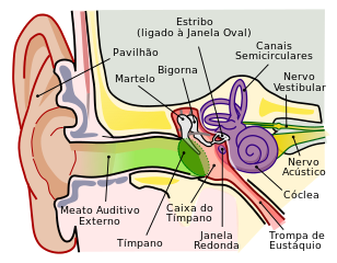
| Description | Anatomia do Ouvido Humano |
| Date | (UTC) |
| Source | File:Anatomy_of_the_Human_Ear.svg |
| Author |
|
| Permission (Reusing this file) |
This file is licensed under the Creative Commons Attribution 2.5 Generic license.
|
| Other versions |
[]
|


| This is a retouched picture, which means that it has been digitally altered from its original version. Modifications: Translated to portuguese. The original can be viewed here: Anatomy of the Human Ear.svg:
|
Licensing
English Wikipedia user Mike.lifeguard, the copyright holder of this work, hereby publishes it under the following license:

|
Permission is granted to copy, distribute and/or modify this document under the terms of the GNU Free Documentation License, Version 1.2 or any later version published by the Free Software Foundation; with no Invariant Sections, no Front-Cover Texts, and no Back-Cover Texts. A copy of the license is included in the section entitled GNU Free Documentation License. |
Original upload log
This image is a derivative work of the following images: *File:Anatomy_of_the_Human_Ear.svg licensed with GFDL, GFDL-user-w **2010-03-09T10:53:44Z Kychot 470x272 (28735 Bytes) {{Information |Source=w:en:File:Anatomy_of_the_Human_Ear.svg |Date=2006-03-06 |Author=w:en:Mike.lifeguard |Permission={{GFDL-user-w|en:wikipedia|English Wikipedia|Mike.lifeguard}} |Category=Anatomy_of_the_ear {{Other versions/Anatomy of the Human Ear}} }} Uploaded with derivativeFX)
File history
Click on a date/time to view the file as it appeared at that time.
| Date/Time | Thumbnail | Dimensions | User | Comment | |
|---|---|---|---|---|---|
| current | 06:55, 11 September 2018 | | 660 × 515 (81 KB) | Jmarchn | Bigger (proportional real size) and full redraw (more realistic) of the auricle. Ossicles in white colour. Eardrum with contour. Added 3 labels. Add fundus to the bone and subcutaneous tissues, add superior auricular muscle, add transparency to middle ear, add separation between middle and inner ear, add division to internal auditory canal. |
| 12:18, 20 May 2010 |  | 800 × 600 (91 KB) | Ruryk | ({{Information |Description={{pt|1=Anatomia do Ouvido Humano}} |Source=Anatomy_of_the_Human_Ear.svg |Author=Chittka L, Brockmann |Date=15 February 2009 |Permission=Creative Commons 2.5|Cathegory=Ear}} ) Category:Ear | |
| 11:59, 20 May 2010 |  | 800 × 600 (91 KB) | Ruryk | Accidental duplication of the original file. The new version contains all labels in Portuguese. Category:Ear | |
| 11:40, 20 May 2010 |  | 800 × 600 (91 KB) | Ruryk | Improvements of some terms. Eliminated one label still in english. | |
| 23:50, 18 May 2010 |  | 800 × 600 (91 KB) | Ruryk | {{Information |Description={{pt|1=Anatomia do Ouvido Humano}} |Source=Anatomy_of_the_Human_Ear.svg |Author=Chittka L, Brockmann |Date=15 February 2009 |Permission= |other_versions= }} |
File usage
The following page uses this file:
Global file usage
The following other wikis use this file:
- Usage on pt.wikipedia.org
- Usage on pt.wikibooks.org